


The Three Phases of Gastric Secretion are:
- The cephalic phase of gastric secretion – takes place as response to stimuli received by the senses
- The gastric phase of gastric secretion – is mediated by the vagus nerve and by the release of gastrin
- The intestinal phase of gastric secretion – involves complex stimulatory and inhibitory process
.jpg)
Cephalic Phase of Gastric Secretion
Food increases gastric secretion through taste pathways when it is present in the mouth. This reflex comes naturally. Additionally, through conditioned reflexes developed during development, the sight, smell, and concept of food trigger stomach secretions. For all these triggers to evoke gastric secretion, efferent vagus nerve should be intact. This is characterized as the cephalic phase because all of the aforementioned stimuli act through higher areas in the CNS.
After three to five minutes of stimulation, stomach secretion begins. It reaches its climax in around 30 minutes, with a 20-minute secretion volume of between 30 and 150 ml.
The entire cephalic phase is an involuntary process that is controlled by the vagus nerve. When food is present or when other senses are stimulated, the vagus nerve, which innervates its branches in the stomach, is activated, causing the stomach's cells to create enzymes and acidic juice.
The postganglionic fibers from the plexus stimulate the chief and parietal cells in various ways, including
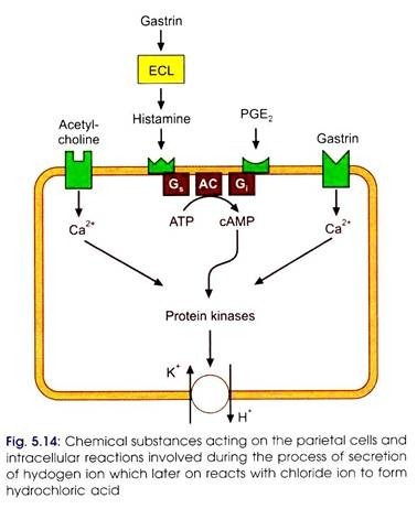
(a) through acetylcholine, which directly affects the gastric glands, and
(b) by stimulating the G cells of the pyloric antrum through Bombesin.
The stomach glands are influenced by gastrin, which is produced by G cells.
Acetylcholine, gastrin, and histamine have distinct receptors on parietal and main cells.
Gastric Phase of Gastric Secretion:
It is also known as the hormonal or chemical phase. The stomach's presence of food boosts gastric secretion. Within 15 minutes of the food entering the stomach, secretion begins. The secretion volume ranges from 225 to 350 ml in 5 hours.
The specific cues that cause this phase are as follows:
a. Mechanical distension of the stomach caused by the food
b. Food derivatives and proteins
These stimuli have a localized effect on the stomach. Their receptors are located in the gastric mucosa, and their efferent local vagal fibers stimulate the ganglion cells in the submucosal plexus, from which the postganglionic fibers stimulate the gastric glands.
Vagus and gastrin both have independent effects on the chief and parietal cells of the stomach, but they also have additive or synergistic effects.
In the pyloric antrum are G cells or APUD cells (Amine precursor uptake and decarboxylation cells). Identical cells are also present in the duodenum.
The hormone that these cells secrete in response to particular stimuli travels through the bloodstream and eventually reaches the stomach glands.
Intestinal Phase of Gastric Secretion:
When stomach material enters the small intestine, the intestinal phase starts. Both stimulatory and inhibitory mechanisms are active during this phase. Although proteins stimulate the production of stomach acid, the chyme prevents this from happening. As a result, the pH of stomach fluids decreases to below 2.5, which prevents the gastrin hormone from being secreted. Thus, only 10% of acid is secreted during this stage.
Proteins and polypeptide stimuli that haven't fully digested are thought to work by causing gastrin to be released in the duodenum. Alcohol can also release duodenal gastrin. When partially digested fat, HCl, and hyperosmolar solutions are present in the duodenum, gastrin release from the pyloric antrum is inhibited, which reduces gastric production.
It is thought that some hormones, such as enterogastrone (a hypothetical hormone), GIP, VIP, Secretin, and glucagon from the duodenum, are responsible for this inhibitory function. Beginning roughly two hours later, the intestinal phase produces 200–300 ml of secretion each hour.
.jpg)
Reference
Guyton AC and Hall JE, 2016. Textbook of Medical Physiology, 13th Edition. Elsevier Saunders, Philadelphia (ISBN: 978-1-4557-7016-8).
Widmaier, EP, Raff, H, and Strang, KT. 2019. Vander’s Human Physiology: The Mechanisms of Body Function, 15th Edition. McGraw-Hill, Boston (ISBN: 978-1-260-08522-8).
R.fb50dfb6ab148ec9541253c256848786 (2143×2775) (bing.com)

.png)

.jpg)
.jpg&description=Discuss the Phases of Gastric Secretion?)
0 Comments